


CS 8/1
Topography of the Orbit
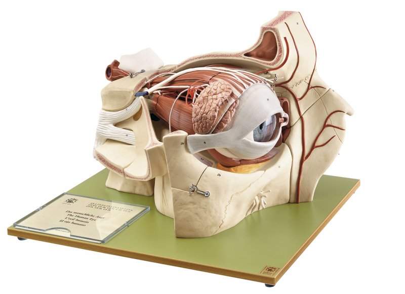
Enlarged approximately 5 times, in SOMSO-PLAST®. The orbital process of the frontal bone and the small wing of the sphenoid bone have been removed in order to allow a view of the bony orbit. The six muscles of the eye are modelled very clearly and the superior and lateral straight muscles of the eyeball can be removed. Separates into 9 parts: Median section of the eyeball (the lens is fixed in the left half), vitreous humour, the right half of sclerotic membrane and choroid membrane with retina can be removed. All important nerves and blood vessels are represented. Lacrimal organs with eyelids. On a green base.
Price on request
Ready to ship today,
Delivery time appr. 1-3 workdays
- Manufacturer number: IDS-6025
- Item number: CS 8/1
- Weight (bw): 8.5 kg
- Height: 33.5 cm
- Width: 45 cm
- Length: 37 cm
- Downloads: Modellbeschreibung (pdf, 0.91 Mb) Model Description (pdf, 1.23 Mb) Descripcion del modelo (pdf, 0.90 Mb)
Viewed